Do Barley’s Trichomes Help Fusarium Head Blight Take Hold? Here’s What New Research Found
Estimated reading time: 4 minutes
By Mitchell Japp, Research & Extension Manager, SaskBarley
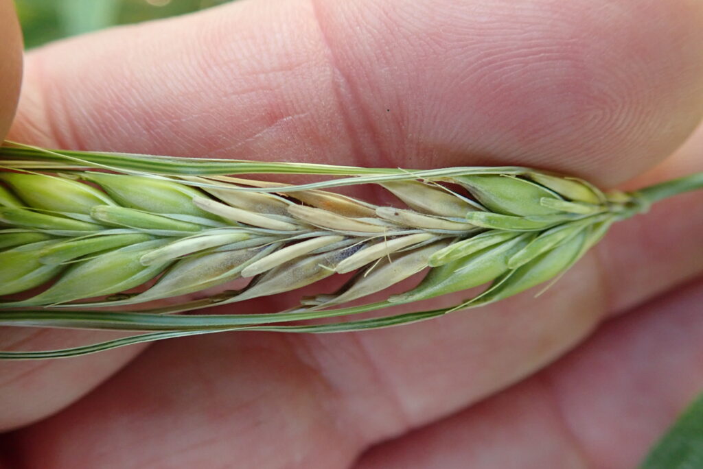
Run your fingers along the awns of a feed barley variety, then a malt variety, and you’ll feel the difference right away — one is smooth, the other grippy or abrasive. That texture comes from trichomes, the bump-, prickle-, spine- and hair-like structures found across barley’s floral tissue. For years, researchers have wondered whether those same structures do more than affect texture: could they be giving Fusarium graminearum, the fungus behind Fusarium head blight (FHB), somewhere to grab hold as it establishes an infection? A newly completed SaskBarley-funded study set out to answer that question directly, and the results reshape where FHB resistance research should look next.
Key Takeaways
- Trichome density varies significantly across barley varieties, tissues and kernel positions, but that variation doesn’t appear to affect how easily Fusarium adheres to the hull.
- Under the microscope, Fusarium spores stuck to smooth surfaces just as readily as trichome-covered ones, sometimes embedding into the hull’s natural waxy coating instead.
- 3D CT scans found a more promising lead: gaps where the two halves of the hull don’t fully fuse together, giving Fusarium a direct route into the kernel.
- The findings rule out trichome density as a useful breeding target, redirecting future research toward wax layer chemistry and hull fusion instead.
Why Researchers Looked at Trichomes
Trichomes have long been suspected of playing a role in how Fusarium establishes itself on barley. Earlier research proposed they might act like tiny hooks — trapping fungal spores and giving hyphae something to anchor to as infection takes hold. If that were true, trichome traits could become a deliberate new target for FHB resistance breeding, something barley breeders don’t currently select for. Dr. Matthew Bakker, of the Department of Microbiology at the University of Manitoba, set out to test that idea directly in a project titled Examining Fusarium Growth and Interactions with Barley Trichomes Under the Hull.
Inside the Research: Electron Microscopes and CT Scans
Bakker’s team collected floral tissue from barley trials and disease nurseries grown in Brandon, Hamiota, Morden, Ottawa and Charlottetown, covering dozens of cultivars and breeding lines. Roughly 150 specimens were imaged using scanning electron microscopy, capturing the inner and outer surfaces of the lemma and palea at the top, middle and bottom of each kernel, along with cross-sections, rachis segments and awns. Because automated counting tools couldn’t reliably handle the complexity of field-grown tissue, trichome densities were counted by hand — painstaking work that built a database of high-resolution images Bakker expects will stay useful to breeders and researchers well beyond this project.
A bonus opportunity let the team push further: using the Canadian Light Source in Saskatoon, they generated computed tomography (CT) scans of dozens of kernels, reconstructing the full three-dimensional interior of each seed at micrometre resolution.
What the Microscopy Revealed
The headline finding runs against the original hypothesis. Barley varieties do differ significantly in trichome density — by variety, by tissue (lemma versus palea), by surface (inner versus outer) and by position on the kernel — but that variation doesn’t translate into a meaningful difference in how well Fusarium sticks around. Fusarium macroconidia and young fungal hyphae adhered to barley hulls just as readily on smooth surfaces as on trichome-dense ones. In some cases, the fungus appeared to embed itself directly into the hull’s natural waxy coating instead, hinting at a different, previously under-appreciated adhesion mechanism.
The CT scans turned up the project’s most promising new lead. Scrolling through cross-sections of individual kernels showed that the lemma and palea — the two halves that fuse together to form the hull — don’t fuse completely everywhere along the seed. At those gaps, Fusarium has a direct, unobstructed route into the kernel. The scans also showed Fusarium-damaged kernels losing much of their aleurone layer, while the large (type A) starch granules inside the endosperm stayed largely intact — suggesting the fungus targets specific tissues and nutrients rather than simply overrunning the whole kernel.
Recommendations for Future Breeding Research
For growers, this project doesn’t change anything in the field today, and that’s a legitimate research outcome in its own right. It rules out trichome density as a promising breeding target, saving future research effort from chasing a trait that likely wouldn’t move the needle on resistance. More usefully, it points researchers toward two new leads worth pursuing: how the natural wax layer on the hull affects fungal adhesion, and how the completeness of lemma-palea fusion might be bred for or managed.
Check out the latest barley varieties with FHB resistance.
Learn more about how breeders are targeting better DON-resistance in new varieties.
PROJECT DETAILS
Project: Examining Fusarium Growth and Interactions with Barley Trichomes Under the Hull
Project Term: March 2021 – April 2023
Principal Investigator: Dr. Matthew Bakker, Department of Microbiology, University of Manitoba
SaskBarley Investment: $50,719
Co-Funder: Manitoba Crop Alliance ($1,568)
Total Project Funding: $52,287